The concept of the compound microscope can trace its roots back to
the late 16th century when the earliest version of a multi-lens
microscope was devised. It was around this time that two notable
Dutchmen, Hans Janssen and his son Zacharias, tinkered with a creation
that would become the forerunner of the compound microscope and the
telescope. It was not until the 17th century, however, that the concept
evolved significantly when a celebrated father of modern microscopy,
Antonie van Leeuwenhoek, crafted microscopes with higher magnification
capability, enabling the discovery of bacteria, spermatozoa, blood
cells, and much more.
Advancements in the 19th century by pioneers such as Carl Zeiss,
Ernst Abbe, and Otto Schott further revolutionized microscopy. They
enhanced optical clarity and illumination, amplifying the microscope’s
power and precision. By the 20th century, electron microscopes were
introduced, achieving revolutionary levels of detail.
The compound microscope, humble in its beginnings, has journeyed
through centuries of scientific refinement and has become one of the
most indispensable tools in today’s scientific and medical fields.
Basic
Definition and Explanation of a Compound Microscope
A compound microscope is an optical instrument designed to make small
objects appear larger through the science of magnification. Named for
their “compound” optical system, comprising two lens systems – the
objective lens and the eyepiece, they provide a detailed view of minute
specimens that the naked eye wouldn’t typically discern.
Interestingly, the term “compound” indicates the use of two or more
lenses working in combination to magnify the image of the specimen. The
first lens, known as the objective lens, magnifies the image initially,
which is then further magnified by the second lens, the eyepiece lens.
By amplifying an object across two stages, the microscope can achieve a
much higher magnification and resolution compared to a simple microscope
which only employs a single lens.
These devices provide a two-dimensional view of the specimen being
observed, with magnification abilities typically spanning from 40x to
1000x, depending on the model in use. They are widely known for their
contribution to various scientific fields, such as biology,
microbiology, and medical research. Moreover, advancements in technology
have given birth to various types of compound microscopes each with
unique features suited to different application needs. However,
regardless of their differences, all compound microscopes share the
quintessential purpose of illuminating the unseen world in vivifying
detail.
Parts of a Compound
Microscope
Overview of Key Parts
Light Source
The light source, often referred to as the illuminator, is located at
the base of the compound microscope. Traditionally, it was a mirror
reflecting ambient light, but modern microscopes typically use a
halogen, LED, or fluorescent light bulb. The primary function of the
light source is to project light upwards onto the slide. This
illuminates the specimen to enhance viewing and allow for better
identification and analysis of the target sample. Some microscopes are
designed with controllable light intensity settings, enabling users to
adjust the brightness level according to the nature and translucency of
the specimen being observed.
Eyepiece
The eyepiece, also known as the ocular lens, is one of the essential
parts of a compound microscope. Positioned at the top of the microscope,
it is the lens you peer through to view the specimen. Typically, the
eyepiece carries a 10x or 15x magnification power, although it can vary
depending on the microscope’s design.
This small, yet crucial lens functions to collect and magnify the
light passing through the objective lens, allowing the viewer to see the
magnified image of the specimen. The eyepiece is often adjustable,
ensuring that users with varying eyesight can clearly observe the
specimen.
In some advanced models of compound microscopes, the eyepiece may
also be equipped with a reticle, or graticule, a type of ruler used for
sizing the objects you are observing.
Objectives
Objectives are an essential part of a compound microscope, located on
a revolving disk known as the nosepiece. They are the lens systems that
focus light into the eyepiece and are used to increase the magnification
of the specimen being examined. Most compound microscopes come with
three or four objective lenses with different magnification powers –
typically 4x (scanning), 10x (low power), 40x (high power), and 100x
(oil immersion objective). Each objective varies in size, focal length,
and magnifying power.
The scanning objective, with the lowest magnification, is used to
provide an overall view of the specimen. The low power is then used for
detailed examination, whereas the high power is used for the most
detailed observations. The oil immersion objective, an interesting and
unique feature of the compound microscope, requires a drop of special
oil placed on the slide, which helps to increase the resolution at the
highest magnifications by reducing light refraction.
Every objective contains a lot of information about its performance
capabilities, etched on the barrel. This could include the magnifying
power, the numerical aperture (a number illustrating the lens’s ability
to gather light), the tube length, and sometimes even the type of
illumination system for which it is designed. By understanding each of
these terms, one can effectively use the objectives to their full
capacity.
Stage
The stage is a crucial component of a compound microscope. It’s a
flat platform where the specimen, generally placed on a glass slide, is
kept for examination. The stage is often equipped with clips or a
mechanical stage to hold and move the slide for viewing different areas
of the specimen.
The stage’s design can vary; however, the majority of compound
microscopes feature a stage that can move up and down, delivering
precise control over the specimen’s distance from the objectives. This
movement allows for accurate focusing. Some modern compound microscopes
also have x and y-axis controls, allowing the user to move the specimen
slide left-right or back-forth without physically touching the slide,
reducing the risk of disturbing the specimen.
The light source is located beneath the stage, shining upward through
the stage’s aperture (an opening or hole). This light illuminates the
specimen from below, enabling the observer to see the specimen through
the eyepiece after it has been magnified by the objectives.
The importance of the stage in a compound microscope cannot be
overstated. It provides the necessary support for the specimen, a
pathway for light, and control over the location and focus on the
specimen.
Function of Each Part
Each part of the compound microscope plays a distinctive role in
order to obtain a detailed and magnified view of the sample.
The light source, typically an LED or a halogen lamp, provides the
necessary illumination for the observation. It directs light upwards
through the sample, aiding in the magnification of the object being
observed.
The eyepiece, sometimes called the ocular lens, is the part that
users look through. It helps further magnify the image formed by the
objective lens, allowing us to clearly see tiny details.
The objectives are a series of lenses with varying magnification
powers. When rotated, they move into place above the sample stage to
magnify the image of the specimen. Low power objectives are used to get
the general layout of the sample, while the high-power objectives
provide a detailed view.
The stage is where the slide containing the specimen is placed for
observation. Some stages come with clips to hold the slide in place and
knobs for precise movement of the slide.
The adjustment knobs, fine and coarse, are used to focus the image.
The coarse adjustment knob quickly brings the object into general focus,
while the fine adjustment knob makes slight modifications for a clear,
sharp image.
In summary, each part of the compound microscope works together to
enhance our ability to observe tiny details that would otherwise remain
hidden to the naked eye.
How a Compound Microscope
Works
The Path of Light
Through the Microscope
A compound microscope does it magic through an intricate dance of
light and lenses. The journey of light within a compound microscope
begins at the light source, typically a bulb beneath the stage that
illuminates the specimen.
From there, the light passes through a condenser, a complex lens
system designed to focus and centralize the light source. After passing
through the condenser, the now-clarified and focused beam of light hits
the slide on which the specimen resides. The light then travels through
the specimen, carrying information about its structure and composition
as it continues its path.
Once the light has passed through the specimen, it encounters the
objective lens, the first of two magnifying lenses. This lens gathers
and magnifies the light, commencing the process of forming an
informative image.
The now magnified light then proceeds upwards via the body tube into
the eyepiece lens, the microscope’s second magnifying lens. This lens
further magnifies and refines the image before it finally reaches the
observer’s eye.
The whole process occurs very quickly and allows us to see
microscopic details of the specimen that are invisible to the naked eye.
Through this combination of carefully arranged light and multiple
lenses, a compound microscope offers a whole new world of exploration
and understanding.
Magnification Process
Magnification is central to the process of using a compound
microscope and involves enlarging the image of the specimen viewed. Each
microscope is equipped with multiple objective lenses with various
magnification powers. The magnification process begins when an image of
the specimen passes through the objective lens, the first one to collect
light from the specimen and form an image.
This image is initially magnified by the objective lens – typically a
4x, 10x, 40x or 100x magnification power. This magnified image is then
viewed through the eyepiece lens, also called the ocular, where it
undergoes another stage of magnification. Common eyepieces magnify the
specimen an additional 10x or 20x.
Final magnification of a specimen can be calculated by multiplying
the magnification power of the objective lens with that of the eyepiece.
For example, if a 10x eyepiece is used in combination with a 40x
objective, the final magnification will be 400x.
It’s important to note that while magnification can make a specimen
appear larger, increasing magnification does not always mean increasing
resolution. Resolution refers to the clarity of the image and depends on
the quality of the lenses and the wavelength of the light used for
observation.
Adjusting the Focus
The focus adjustment on a compound microscope is a pivotal aspect of
its functioning, ensuring the clarity and brightness of the image being
viewed. It typically involves two knobs – the coarse adjustment knob and
the fine adjustment knob.
The coarse adjustment knob is used initially to bring the specimen
into the range of focus, rapidly moving the stage up and down. Once the
image is roughly in focus, the fine adjustment knob is used to bring the
specimen into sharp focus by making slight alterations to the stage’s
position.
Focus adjustment is generally performed under low power before
switching to the higher power objectives. It’s important to adjust the
focus incrementally and with care to avoid damage to both the lens and
the specimen, as very high power objectives might touch the slide if not
carefully adjusted. Thus, mastering the skill of focus adjustment is
essential for maximising the effectiveness of a compound microscope.
Types of Compound
Microscopes
Monocular Compound
Microscope
A monocular compound microscope is perhaps the most recognizable
variant of the compound microscope series, especially in educational
settings. As the name suggests, ‘monocular’ means one, implying this
microscope design has a single eyepiece for observation. Despite the
presence of a singular eyepiece, it still utilizes the classic compound
system involving multiple lenses to maximize magnification.
One significant feature of the monocular compound microscope lies in
its affordability and portability, making them popular for use in
classrooms and fieldwork. Furthermore, these microscopes are generally
simpler to use, since users only view specimens through one eye,
reducing the potential for eye strain or discomfort over long periods of
use.
One should note that while they may lack the increased depth
perception found in binocular models due to its singular eyepiece,
monocular compound microscopes still provide ample detailing and
magnification for most general microscopy needs. Their compact design
and simplicity of use make them an essential tool in the world of
optical magnification.
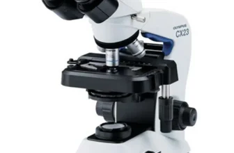
Binocular Compound
Microscope
Binocular compound microscopes incorporate two eyepieces, as their
name suggests, compared to the single eyepiece in monocular microscopes.
This quality doesn’t necessarily give a stereoscopic, or 3D vision, as
one might assume, but it does provide a significant advantage: the
ability to view samples for extended periods without causing intense eye
strain, thus extending the duration of potential research and study.
The two eyepieces on a binocular compound microscope are designed to
observe the same image, each working accessories to a straight eye-level
tube. The user can adjust the inter-pupillary distance (the distance
between the centers of the pupils of the eyes) to view the specimen
comfortably.
This type of microscope is immensely popular among serious
researchers and professionals as it facilitates long hours of detailed
study. The twin eyepieces distribute the optical load evenly, reducing
eye fatigue. Furthermore, a degree of depth perception is maintained,
providing a generous and comfortable field of view for users who wear
glasses. An additional advantage is the eyepiece’s ability to rotate,
allowing multiple viewers to simultaneously observe a specimen from
different angles without adjusting the instrument’s position.
Trinocular Compound
Microscopes
Trinocular compound microscopes are similar to their binocular
counterparts, but with an added feature that sets them apart. These
microscopes have three eyepieces, thus their name ‘trinocular.’ The
third eyepiece usually doesn’t contribute to the direct observation
process, but instead is often used for camera attachments. The main
advantage of this type is that it allows for real-time viewing and
recording of the microscope’s observations. It provides the ability to
capture, share, or store the images digitally. Scientists, researchers,
and educators frequently use trinocular microscopes to document their
findings and to aid in collaborative work or instruction. These
microscopes are especially prevalent in fields that require precise
documentation such as microbiology, medicine, and forensic science.
Applications of Compound
Microscopes
Use in Scientific Research
Compound microscopes have played a crucial role in advancing our
understanding of the world around us. They are integral to various
fields of scientific research, including microbiology, genetics,
biochemistry, and pharmacology, to name a few.
In microbiology, compound microscopes are used to inspect bacterial
cultures. By magnifying the sample, scientists can identify specific
bacterial species based on their shape and staining properties, giving
insights into the root of infections or diseases.
Genetics also relies heavily on compound microscopes. The detailed
view provided by the microscope allows scientists to observe and analyze
chromosomes – DNA carriers – in cells. This has led to significant
breakthroughs in our understanding of hereditary conditions and genetic
diseases.
Another field, biochemistry, uses compound microscopes to analyze the
reactions of organisms to various chemical compounds. This is
fundamental in the development of new drugs and treatments.
Similarly, in pharmacology, compound microscopes help validate the
effect of drugs at a microscopic level. They are used to monitor the
cellular responses to various drug treatments, aiding in the design of
more effective therapeutic agents.
In short, compound microscopes play an indispensable role in
scientific research. Leveraging their high magnification and resolution
capabilities, they enable scientists to observe and understand actions
and reactions that are otherwise invisible to the naked eye. The
information gleaned from microscope studies provides the foundation for
theories, solutions, and advancements that propel our scientific
landscape forward.
Use in Education
High School Biology
High school biology is one of the primary places where compound
microscopes play an essential role. At this stage of education, students
begin to delve into the microscopic world, exploring the cellular
structure of plants and animals.
A compound microscope is an invaluable tool, providing students with
hands-on experience as they study different cell types, observe cellular
division, and examine various fibers, tissues, and other microorganisms.
It offers students the possibility to explore beyond what can be seen by
the naked eye and build a foundation for understanding concepts like
cell theory, anatomy, and microbiology.
Moreover, using a compound microscope in high school biology can
foster critical thinking and problem-solving skills. It enables students
to observe and analyze microscopic specimens, test hypotheses, and reach
data-driven conclusions. This element of practical investigation forms a
significant part of the curriculum and helps students develop scientific
literacy.
By using compound microscopes, students get the first taste of what
it is like to be a scientist, observing the minute details of life
unseen by the naked eye. They become active participants in the learning
process, transforming abstract concepts into concrete understanding.
This interaction with the microscopic world might inspire some students
to pursue further studies or careers in life sciences and related
fields.
In summary, compound microscopes serve as a window into a fascinating
world of minute life occurrences, shaping the educational journey of
high school biology students and bringing the textbook to life.
College-Level Microbiology
Compound microscopes play a vital role in college-level microbiology,
shaping the educational experience for students who delve into the
microscopic world of microorganisms.
In microbiology laboratories, microscopes are routinely used to
examine the structure, functions, and behavior of various
microorganisms. It allows students to identify bacteria, viruses, and
fungi, based on their physical and biochemical properties. By observing
slides stained with different dyes, students can visualize cellular
structures that would otherwise remain invisible to the naked eye.
Furthermore, microbiology students use compound microscopes for
numerous laboratory methods, including Gram staining and cell culture
analysis, enabling them to identify and classify pathogenic
microorganisms. Additionally, compound microscopes aid in the
understanding of microbial genetics, providing a visual aid when
analyzing replication, gene expression, and genetic mutation in
microorganisms.
For aspiring microbiologists, familiarity with compound microscopes
during their college education is crucial. This tool helps not only in
developing their technical skills, but also in understanding the
intricate details of microbiology, thus expanding their knowledge in the
field.
In conclusion, the role of compound microscopy in the realm of
college-level microbiology is undeniably crucial, as it acts as a
gateway to understanding the vast world of microorganisms, driving
education and research forward.
Use in Medical Diagnosis
Compound microscopes play a crucial role in medical diagnosis. They
aid in the visualization of cells and tissue structures in great detail,
which is incredibly vital in identifying diseases at a cellular level.
They are used in a plethora of diagnostic procedures, including analysis
of blood samples, tissue biopsies, and microbial cultures.
In a blood test, for instance, a compound microscope helps in
counting and assessing the shape, size, and overall condition of
different blood cells like red blood cells, white blood cells, and
platelets. Changes in these factors can reveal diseases such as anemia,
leukemia, malaria, and various immune disorders.
Moreover, compound microscopes provide the ability to observe tissue
samples in histopathology. Pathologists examine the biopsied tissue to
look for abnormalities, which can indicate a variety of conditions,
including cancer. Different types of stains are used to highlight tissue
structures, cells, and cell components, making it easier to spot
anomalies.
Additionally, compound microscopes are indispensable in microbiology
laboratories. They allow for the identification and characterization of
bacteria, viruses, fungi, and parasites. This is particularly essential
for the diagnosis of infectious diseases and deciding on the appropriate
course of therapy.
Thus, the use of compound microscopes in medical diagnosis is vast,
making it a crucial tool in the healthcare industry.
Advantages
and Disadvantages of Compound Microscopes
Benefits of Using
Compound Microscopes
Compound microscopes present a plethora of benefits that have made
them essential tools in various fields. First off, they enable users to
observe the intricate details of minute objects, unveiling an unseen
world that otherwise remains invisible to the naked eye. From bacteria
to the fine-grained structure of materials, these microscopes offer a
deep dive into the microcosm of life and matter.
They provide significant magnification, often up to 1000x or more,
which makes them incredibly effective for biological research. This is
particularly beneficial for studies involving cells, microorganisms, and
small species. Another advantage lies in their ability to produce a
3-dimensional image of the specimen, contributing towards a more
comprehensive understanding of its structure.
Furthermore, compound microscopes provide a high level of contrast,
making it possible to differentiate between different parts of a sample
or between different organisms in a mix. They can also be used with a
variety of staining techniques, making certain features stand out more
clearly.
One of the most crucial benefits of compound microscopes is their
versatility. Variations of compound microscopes, such as
stereomicroscopes or confocal microscopes, cater to diversified needs
and applications. Lastly, considering their function, compound
microscopes are reasonably affordable, making them accessible to
educational institutions, research centers, and hobbyists alike. Even
high-end models, while pricier, provide superior optical quality and
extra features that are invaluable for certain types of research,
validating their cost.
In conclusion, the application of compound microscopes transcends
across fields, expanding the dimensions of what we can explore, study,
and ultimately understand. Despite certain limitations, the benefits
they offer significantly outweigh these, asserting their indispensable
role in science and technology.
Limitations of Compound
Microscopes
While compound microscopes boast numerous advantages, they are not
without limitations, and careful consideration to these shortcomings is
essential depending on the purpose they are intended to serve.
One of the most significant limitations of compound microscopes is
their restricted resolution. The phenomenon of diffraction restricts
further improvement in resolution under normal conditions, often making
it challenging to discern detail in very small specimens. Therefore,
compound microscopes might not be suitable for observing features
smaller than approximately 0.2 micrometers.
Another limitation is the two-dimensional depth of field. While this
is beneficial for studying flat samples, it becomes a hindrance when
examining specimens that require depth perception. Thus, for larger, 3D
specimens, a compound microscope may not provide a comprehensive
view.
Also, the need for perfect specimen preparation is another drawback.
The samples have to be sliced into extremely thin segments, and certain
substances might not lend themselves to this kind of preparation.
Besides, some preparation processes like staining and fixing may alter
the actual properties or behavior of the specimen being observed.
Lastly, most compound microscopes are not portable due to their
weight and size. This can limit their usage in certain field situations
where carrying heavy equipment is impractical.
Keep in mind that while these limitations exist, the compound
microscope remains a valuable tool, thanks to its versatility and
ability to provide a magnified view of speciments. Understanding these
limitations is essential for picking the right tool for specific
microscopic needs.
Conclusion
Reiteration
of Microscope’s Importance and Relevance
Throughout this article, we have learned the ins and outs of the
compound microscope – from its humble beginnings to its modern-day
incarnation. It’s clear that this device holds a sense of timeless
relevance and importance across various fields. It is thanks to these
complex instruments that scientists, students and medical professionals
are able to explore, study and understand the minutiae that lie beyond
our ordinary vision. This intimate look into the microscopic world not
only enhances scientific research, medical diagnostics, and education
but also opens up doorways to groundbreaking discoveries in all corners
of science. Any progress in our society today, be it in healthcare or in
our understanding of the world, owes some credit to the compound
microscope. As we forge ahead into the future, the compound microscope
is sure to remain an indispensable tool for exploration and
discovery.
Final
Thoughts on the Evolution and Future of Compound Microscopes
Compound microscopes, since their inception, have played a pivotal
role in revolutionizing our understanding of the microscopic world.
Their evolution from rudimentary magnifying tools to sophisticated
pieces of scientific equipment speaks volumes about the progress of
scientific technology and our unrelenting quest for knowledge.
Looking to the future, it is expected that compound microscopes will
continue to evolve with advancing technologies. Developments in areas
like software enhancements, digital imaging, and nanotechnology will
potentially offer more powerful and precise imaging capabilities.
Innovations in areas like automating slide movement, advanced
staining procedures, and image analysis software will likely increase
efficiency and accuracy of microscopy-based research and diagnosis.
Additionally, the integration of artificial intelligence could help in
automated identification and classification of microscopic entities,
which could revolutionize fields like pathology.
Therefore, the future of compound microscopes seems exciting,
promising to break new boundaries in our exploration and understanding
of the microscopic world. Their ongoing evolution assures us that these
invaluable instruments will remain an essential part of scientific
advancement, maintaining a vital role in research, education, and
medical diagnosis for years to come.